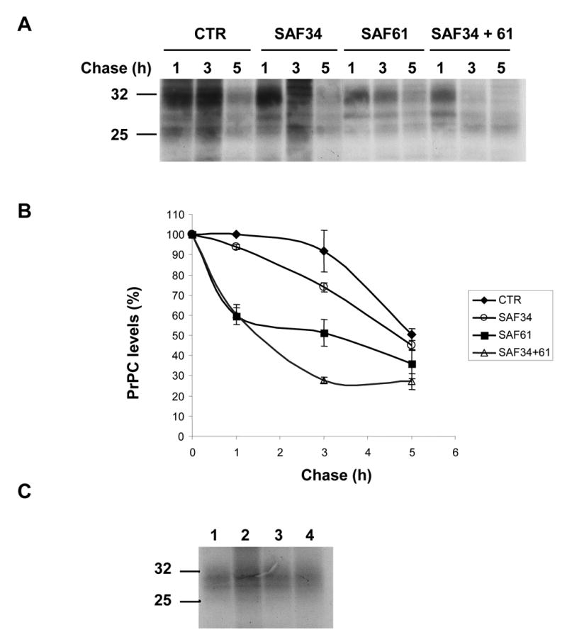
Figure 5. Anti-PrP antibodies decreased the half-life of the PrPC protein
(A) Non infected N2a58 cells in 35 mm dish were pulse-labeled for 1 h in absence of anti-PrP antibodies. During the chase of 1, 3 and 5 hours, cells were incubated with 2 μg/ml of anti-PrP antibodies, either SAF34 or SAF61 or both of them. At the end of the chase, PrPC proteins from the cell lysates were immunoprecipitated according to the protocol described in Mat. & Meth. Samples were then loaded onto a 12.5 % SDS-PAGE gel, followed by an autoradiography. (B) Densitometry analysis of three autoradiographies were done to determine the half-life of the PrPC proteins. PrPC bands were quantified using the image analysis software Scions Image Beta 4.02. The maximum normalized value corresponds to the control cells radio-labeled during 1h, with no chase period and without antibodies added in the medium. For each PrPC band, we subtracted a background value, selected in their vicinity. All the densitometry values were normalized in comparison with the control. Data represents the mean from 3 independent experiments, with errors corresponding to the standard deviation. (C) Non infected N2a58 cells in 35 mm dish were pulse-labeled for 30 min and simultaneously incubated with 2 μg/ml of anti- PrP antibodies. Control sample without anti-PrP antibodies (1); cells incubated with either SAF34 (2), or SAF61(3) or both of them (4).