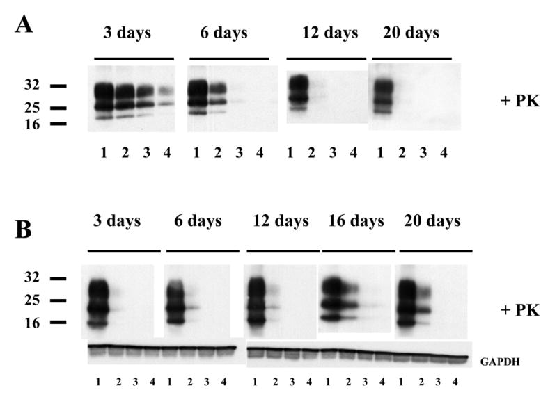
Figure 3. Disappearance of PrPSc molecules from prion-infected N2a58/22L cells treated with the purified antibodies
(A) Cells cultured in 25-cm2 flasks in the presence of purified antibodies were serially passaged every 3 days. At each 3 days split, antibodies were freshly added to the medium. PrPSc molecules were detected in cell lysates previously digested with proteinase K according to the protocol described in Material & Methods. (1) Control without treatment with the antibody; (2) Cells treated with 1 μg/ml of SAF34; (3) Cells treated with 1 μg/ml of SAF61; (4) Cells treated with 1 μg/ml of SAF34 and 1 μg/ml of SAF61. (B) Cells that were previously treated for 20 days with the antibodies were then cultivated in the absence of antibodies, for another 3 to 20 days period. Cell lysates were digested with proteinase K to detect the PrPSc isoform. Molecular weight markers spanning 16.5–32 kDa. Immunoblots were probed with SAF mix antibodies. The immunoblots selected in this figure are representative of 3 independent experiments.