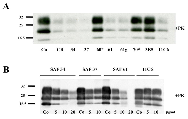
Figure 1. Screening of monoclonal antibodies in prion-infected N2a58/22L cells
(A) PrPSc levels were analyzed by immunoblotting after 3 days of culture in presence of non-purified antibodies SAF34, SAF37, SAF60, SAF61, SAF61g, SAF70, 3B5 and 11C6 at a concentration of 10 μg/ml. Co is the level of PrPSc without antibody treatment. CR is the positive control for inhibition of PrPSc, after treatment of the cells with 1μg/ml of Congo red. (B) Dose-dependent inhibition of PrPSc formation with non-purified antibodies SAF34, SAF37, SAF61 and 11C6. Cells were incubated with various concentration of antibodies (5 to 20 μg/ml) for 3 days. Samples were digested by 20 μg/ml of proteinase K at a ratio of 1:50 protease to protein for 1 hour at 37°C. PrPSc levels were analyzed by immunobloting. Molecular weight markers spanning 16.5–32 kDa. The star symbol means that the antibodies were not able to immunoprecipitate the PrP protein (see Table 1). Immunoblots are probed with SAF mix antibodies.