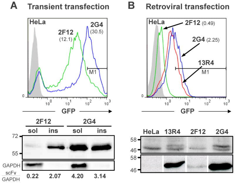
Fig. 2.
Cytoplasmic expression and whole-cell fluorescence of EGFP tagged 2F12 and 2G4 intrabodies. A) HeLa cells were analysed 24h after transient transfection with plasmid pCMV-SN-EGFP containing the indicated scFv. Top: FACS analysis using the GFP fluorescence signal. The geometric means of the GFP+ cells are 12.1 for 2F12 and 30.5 for 2G4. The region M1 identified by the horizontal line contains 40% of the 2G4 and 10% of the 2F12 population. Bottom: Western blot of detergent-soluble and -insoluble fractions of the same cell populations. The fractions from 150,000 cells were analysed in each lane. The blot was revealed and quantified as in Fig. 1. B) HeLa cells were infected with retrovirus containing 2F12-GFP, 13R4-GFP and 2G4-GFP fusions and the cell populations analysed by: Top: FACS using the GFP fluorescence signal. The geometric means of the GFP+ cell population are, respectively, 0.49 and 2.25 for 2F12 and 2G4. The region M1 contains 40% of the 2G4 and 0.1% of the 2F12 population. Bottom: Western blotting of; Top-Panel: Total cell extracts revealed using a rabbit polyclonal anti-GFP serum; Bottom-Panel: scFv-GFP fusions were captured from soluble cell extracts by their C-terminal polyHis tag and detected using the 9E10 anti-c-myc monoclonal antibody. In all cases, lanes are from the same gel but were reordered to help the reader.